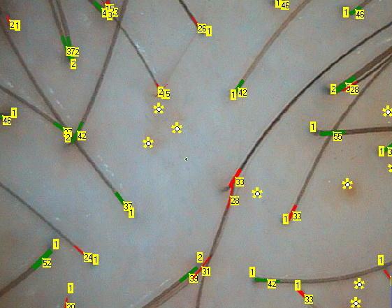
Trichoscopy, as a method of assessing a condition of hairs of a scalp, eyebrows and eyelashes with a video- dermatoscopic technology, was first introduced by Lidia Rudnicka in 2006. Trichoscopy module is a complex tool, which contains separate "General Trichoscopy", "Hair diameter" under higher magnification, "Scalp analysis", "Hair roots analysis" and "Hair shafts analysis" diagnostic sessions. General Trichoscopy section allows to estimate hair density and diameters in different zones of the scalp, as well as to access hairs distribution in follicular units and perifollicular sign counts, while hair diameters can be calbrated more precisely under higher maginification in spearate sections, if necessary. Scalp, hair roots and shafts analysis diagnostic sections include an appropriate sample image databases to aid in a proper preventive diagnosis establishent. General Trichoscopy section for hair density counts simultaneously with hairs diameters measurements. Measurements can be carried out in a semi-automatic or manual modes. Other functions include mark ups and counts for "Follicular unit" with a number of hairs in each and “Perifollicular sign”, including “Pointed hair”, “Exclamation mark hair”, “Broken hair”, “Cadaverized hair”, "Yellow dots", "Red dots" and "White dots“. “Linear length” function allows to perform hair length measurements of any growing hair within the site of view. "Point localization "function allows to mark up specific measurement points on scalp diagram, where hair counts have been performed.
In addition to hair density and diameters evaluation, the severity of Anisotrichosis (or Polymorphism, that reflects the degree of deviation of hair diameters from norm), which is an important parameter that assesses progressive hair thinning, is taken into consideration along with the percentages of Vellus-like hair less than 30 microns in diameter. This allows for a more comprehensive evaluation of the severity of ongoing pattern alopecia processes. In these cases it is also quite important that hair assessments are not limited to diameter estimates only, but include classification by type (i.e. thin, medium and thick hair) along with calculations of the percentages for each of those types of hair. The resulting data is important for assessing a current hairs condition, as well as for a dynamic observation of patients during treatment or scientific studies. In each field of view, it is recommended to account for a presence of various Perifollicular signs, such as ”yellow dots” (reflect delays of new hair growth phases), “white dots” (reflect a presence of follicle fibrosis, typical for scarring forms of alopecia), “spiky hair” (reflect an intensity of hair loss), “red dots” (reflect vascular changes, typical for Psoriasis, Discoid Lupus), hair in a form of an “exclamation mark” and“ black dots” (characteristics of Alopecia Areata).
Hair diameter measurements and subsequent evaluation can be carried out under higher magnification, thus, allowing for a greater accuracy while obtaining data. Measurements can be carried out in a semi-automatic or manual modes. A proper assessment of a scalp condition enables to detect changes in the perifollicular zone, that shall be considered when selecting a treatment for patients with alopecia and/or dermatosis signs. A proper microscopic evaluation of extracted hair roots allows to quickly and accurately differentiate Anagen Alopecia from Telogen Alopecia. For example, presence of more than 80% of dystrophic hair roots in Anagen phase is a characteristic of Anagen Alopecia, which is associated with influence of toxic factors, or autoimmune reactions. Dystrophic hair has a shattered bulb, conically narrowing shaft and no root sheath. In dysplastic hair root bulb is deformed, reduced in diameter, root sheath is completely or partially absent. Dysplastic and dystrophic hairs typically represent signs of Alopecia Areata, however, may also be present in hair loss induced by factors, which affect hair follicle state at dermal papilla, such as effects of chemo or radiation therapy, poisoning by salts of heavy metals, due to anticoagulant or interferon medication therapy, etc. While microscopy of hair shafts allows to reveal various defects of hair keratinization, that are hereditary in nature, as well as hair structural damages associated with improper care due to cumulative effects of physical, chemical and mechanical actions.
For more Trichoscopy module features of the TrichoSciencePro © computer program refer to Chapters 3 to 11 of the User Manual. In case of any recent updates, for some additional information please also refer to the publication for the New Version.